FTIR microscopy

Applications for polymers

FTIR microscopy (µFTIR) combines infrared spectroscopy with a microscopic setup to investigate the chemical composition of polymers with spatial resolution. µFTIR provides detailed chemical imaging of small sample areas, identifying the distribution of specific chemical bonds and functional groups throughout the sample. It is a powerful technique to determine different phases or domains within a polymer matrix (polymer blends, copolymers, additives, impurities, degradation products, composites, multilayer films). Therefore, µFTIR has great potential to answer current questions for R&D and Q&S of plastics and plastic products.

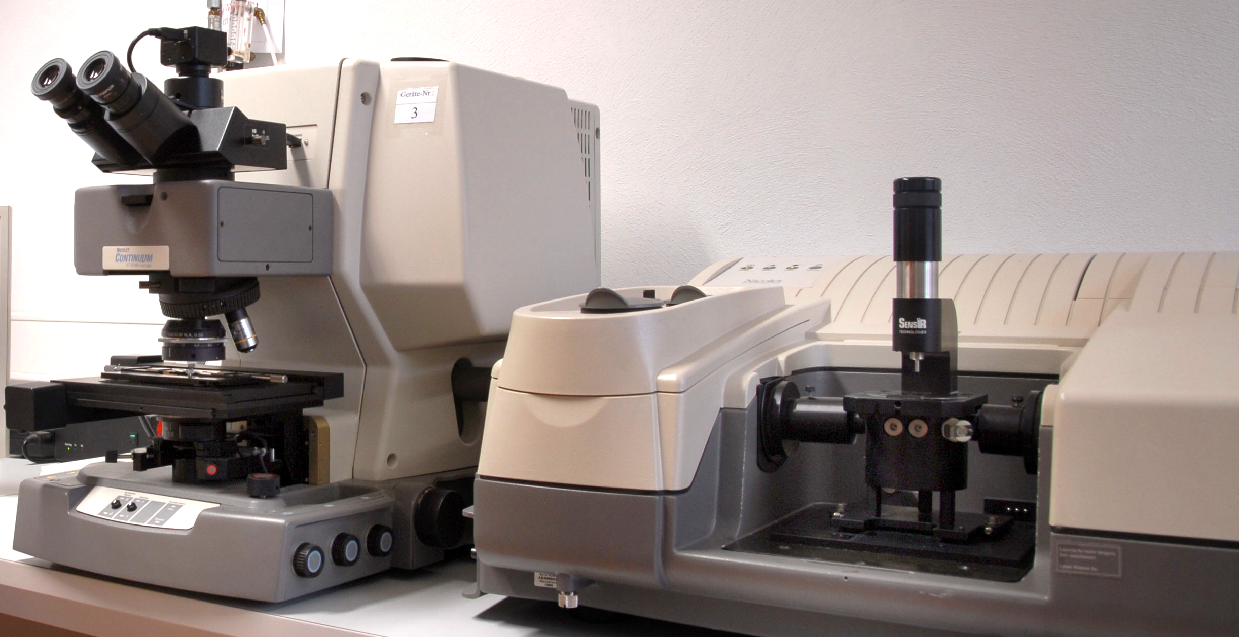
FTIR microscope Thermo Nicolet Continuum with connected FTIR spectrometer Thermo Nicolet 8700 at Fraunhofer LBF

Use cases

Additive migration

  • Tracking the stabilizer concentration in hot water pressure pipes made of different PE and PP types during accelerated ageing at different temperatures and pressures

Figure:

During accelerated ageing at 80 °C in the creep-rupture internal pressure test, the stabilizer concentration in PE 100 decreases more slowly than in PE80, despite the higher internal pressure for PE 100 (11 bar) compared to PE 80 (10 bar). This behavior, which was monitored by FTIR microscopy, indicates a higher long-term resistance of PE 100.

Root-cause analysis

  • Analysis of damaging effects in domestic water installations made of PP caused by different types of disinfectants (chlorine gas, chlorine dioxide)

Figure:

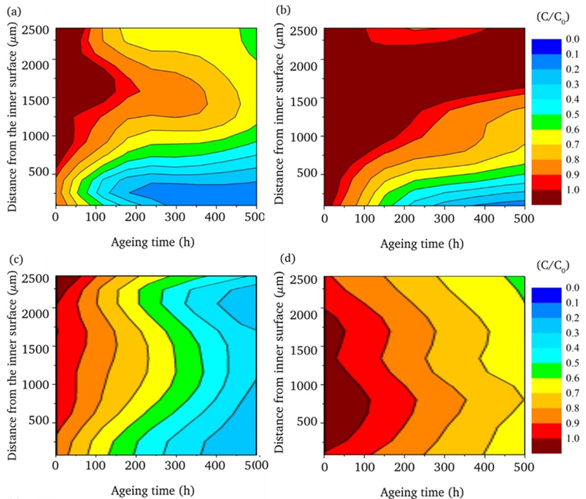
Chlorine-containing disinfectant (a, b) reduces the stabilizer concentration on the inner wall of the pipes considerably faster during accelerated ageing at 95 °C than water (c, d). The PP grade with increased long-term resistance shows a slower decrease in stabilizer concentration than the standard PP grade (a, c) both in chlorine-containing disinfectant (b) and in water (d). If the stabilizer concentration falls locally below a critical limit value (e.g. 10 % of the initial concentration), slow crack growth is initiated.

Quality assurance

  • Use of nucleating agents to improve the quality of weld seams in PP injection molded parts

Figure:

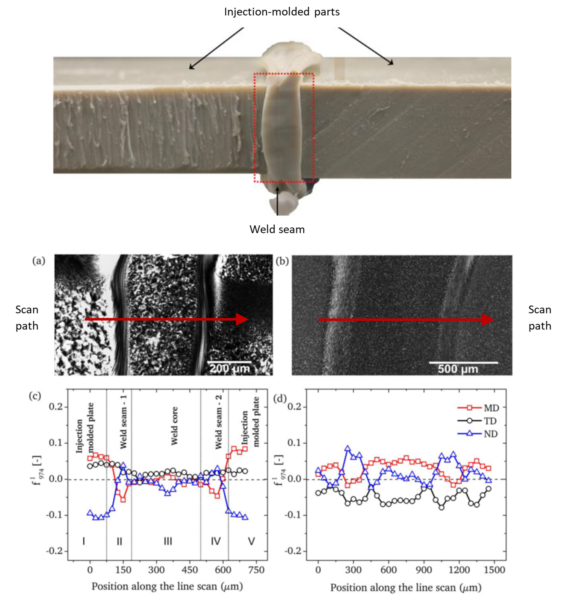
Dichroism measurements using FTIR microscopy reveal the effects of nucleating agents on the quality of polypropylene weld seams.
Top image: Injection-molded parts joined by butt welding.

  • a) Polarized light microscopy (PLM) image of a thin section from the welded area of a conventional polypropylene (PP) material, showing the semicrystalline morphology and the heat-affected zones resulting from the welding process.
  • b) Polarized light microscopy image of a thin section from the welded area of a specialized polypropylene (PP) material equipped with nucleating agents. Compared to conventional PP (a), this results in a finer semicrystalline morphology and less pronounced heat-affected zones under identical welding parameters.
  • c) Degree of dichroism along a measurement path taken in different sectional planes relative to the weld seam in (a) (MD, TD, ND). The degree of dichroism is a measure of the orientation of polymer molecules within the crystalline domains (spherulites) of the PP material. A preferred orientation of the polymer chains in the MD and ND directions (biaxial orientation) is evident. The local progression of the dichroism degree can be correlated with the morphological features seen in the PLM image from (a). Accordingly, molecular orientation within the weld seam is only weakly pronounced and increases abruptly toward the edges. Such abrupt changes in molecular orientation (singularities) are expected to reduce the weld seam strength.
  • d) Degree of dichroism along a measurement path taken in different sectional planes relative to the weld seam in (b) (MD, TD, ND). No preferred orientation of the polymer chains is observed. The local progression of the degree of dichroism can be correlated with the morphological features seen in the PLM image from (b). Accordingly, the molecular orientation inside and outside the weld seam is very similar, with no abrupt increase toward the edge. The uniform morphology of the weld seam is expected to result in higher strength of the nucleated PP material compared to conventional PP.

Process Analytical Technology

  • Ex-situ tracking of the aging behavior of hot water pressure pipes made of PP-R with different extrusion speeds.

Figure:

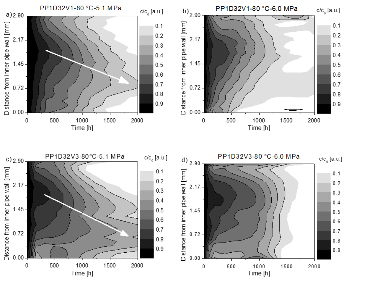
Concentration of the phenolic stabilizer Irganox 1010 as a function of location and time during the long-term internal pressure test of hot water pressure pipes made of polypropylene. PP pipes extruded at low (a, b) and high speeds (c, d) were tested. The dwell time of the stabilizer is greater in the PP pipe extruded at higher speeds (white arrow).

Techniques

  • Transmission IR spectroscopy: A thin polymer film is exposed to IR radiation and the transmitted light is recorded. A detailed spectrum is obtained, but due to the thickness of the sample, it may be necessary to prepare a thin section on the microtome. Transmission IR microscopy offers an advantage in the quantification of components in polymer samples, as the prepared sample thickness can be precisely controlled. This allows trace components such as additives to be detected at concentrations as low as 200 ppm.
  • Attenuated total reflection (µATR): This method uses a tip to internally reflect the IR beam that is in contact with the sample (contact mode). This allows directly analyzing samples at their surface without special sample preparation. µATR-IR is particularly useful for thicker and uneven samples.
  • Transmission-reflection IR microscopy (transflection): This method is used to examine thin films that are applied to metallic surfaces. For this purpose, the infrared beam is directed onto the sample surface in reflection mode. The radiation is reflected by the metallic substrate and analyzed in the detector after passing through the film twice.

Basics

Infrared light interacts with the sample and causes molecular vibrations that are specific to different chemical bonds and functional groups. These vibrations lead to absorption peaks at characteristic wavelengths (FTIR spectrum), which is generated in localized form from small areas of a sample. FTIR microscopes are often coupled with FTIR spectrometers, which serve as a source for the modulated infrared light. They are equipped with a sample stage that holds and positions the sample for analysis. The objectives of the microscope focus the infrared light on a small area of the sample and collect the transmitted or reflected light. The infrared light that has interacted with the sample is captured by the detector.

Publications:

  • Maria, R., et al., Ageing study of different types of long-term pressure tested PE pipes by IR-microscopy, Polymer 61 (2015), 131-139, https://doi.org/10.1016/j.polymer.2015.01.062
  • Damodaran, S., et al., Measuring the orientation of chains in polypropylene welds by infrared microscopy: A tool to understand the impact of thermo-mechanical treatment and processing, Polymer 60 (2015), 125-136, https://doi.org/10.1016/j.polymer.2015.01.046
  • Schuster, T., et al., Quantification of highly oriented nucleating agent in PP-R by IR-microscopy, polarised light microscopy, differential scanning calorimetry and nuclear magnetic resonance spectroscopy, Polymer 55 (2014), 1724-1736, https://doi.org/10.1016/j.polymer.2014.02.031
  • Geertz, G., et al., Stabiliser diffusion in long-term pressure tested polypropylene pipes analysed by IR microscopy, Polymer Degradation and Stability 94 (2009), 1092-1102, https://doi.org/10.1016/j.polymdegradstab.2009.03.020